
Evaluation of eyelid sagging with Antera 3D by Miravex via FOCUS#12
26 June 2025
The Antera 3D system is a high-resolution, non-invasive imaging device used to analyze skin properties in 3D, particularly helpful in assessing pigmentation, vascularity, texture, and skin surface topography. Although the Antera 3D doesn’t measure mechanical properties – e.g. elasticity, tensile strength – directly, it can complement biomechanical assessments by:
- Documenting surface-level changes following interventions aimed at improving biomechanical properties (e.g., after microneedling, radiofrequency, or topical retinoids).
- Helping correlate surface structure with elasticity changes measured by other instruments.
Here we report a study carried out by Hurley S. et al. [1] on the assessment of eyelid sagging.
As the eye contour ages, the skin on the lid becomes lax often causing a voluminous protrusion where the superior palpebral sulcus begins to sag onto the upper eyelid. To investigate the possibility of quantifying the volume of the sagging, eyelid topographic measurements were collected on 20 female volunteers aged 50‐75 years and the volume evaluated with the DermaTOP (Eotech) and Antera 3D (Miravex).
Figure 1 shows the Volume (mm3) of the sagging feature ± SD at baseline and 5 minute follow-up, on the same eye, without treatment. No significant change in feature volume with regard to baseline measurement was recorded using either the DermaTOP or the Antera 3D data.
[1] Hurley S. et al., “DermaTOP Blue and Antera 3D as methods to assess cosmetic solutions targeting eyelid sagging”, Skin Res Technol. 2019;00:1–6.
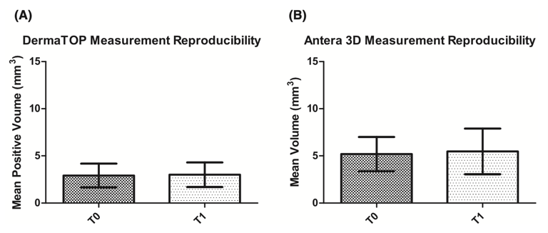
Figure 2 shows the mean Volume (mm3) of the sagging feature ± SD at baseline and 10 minute follow‐up, on the same eye, after a single application of an aqueous tightening serum for the DermaTOP (2A) and 5 minute follow‐up for the Antera 3D (2B). A significant 12% reduction in feature volume with regard to baseline measurement was recorded using the DermaTOP data and a significant 21.6% reduction in feature volume was recorded using the Antera 3D.
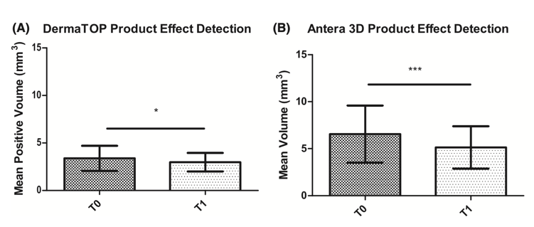
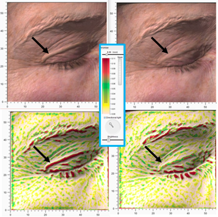
Figure 3 shows Antera 3D Elevations Channel (Custom 1.1 mm filter) capture for a volunteer at baseline and 5 minutes after a single application of aqueous tightening serum on the eyelid area.
This study has demonstrated that both the DermaTOP and Antera 3D allow for quantitative measurement of eyelid sagging feature volume and in‐turn permit evaluation of anti‐ageing cosmetic preparations targeting eyelid sagging.
CONTACT
Guido Mariotto
CEO
guido@miravex.com
https://miravex.com/
https://www.skinobs.com/c/manufacturer.php?id=72


 Follow us on Linkedin!
You must be logged in to post a comment.