A Model To Study Dermal Cells Contractility by Syntivia
29 July 2024
ROLE OF DERMAL FIBROBLAST CONTRACTILITY IN THE SKIN
The contractility of dermal fibroblasts plays a crucial role in the skin, particularly in the processes of wound healing and tissue repair, but also in skin aging.
During the healing process, fibroblasts migrate to the site of injury. They are responsible for producing new tissue by synthesizing extracellular matrix components. The cells’ contractility enables them to remodel skin tissue for better healing, by adjusting tissue tension and optimizing the strength and flexibility of the repaired skin.
Beyond wound healing, fibroblast contractility is important for maintaining skin tension and elasticity, which is essential for its barrier function and for preserving the skin’s youthful appearance. Aged fibroblasts show an altered capacity to contract.
DERMAL FIBROBLASTS CONTRACTILITY ASSAY
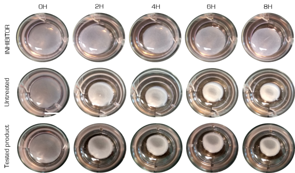
Syntivia has validated a method for assessing fibroblast contractility using collagen gels incorporated with fibroblasts to evaluate the contractile capacity of the cells. After polymerization of the lattice, fibroblasts reorganize and contract the collagen network. Syntivia is working on Normal Human Dermal Fibroblasts (NHDF) from any type of donor seeded in a collagen gel (figure 1).

Analysis of contraction power
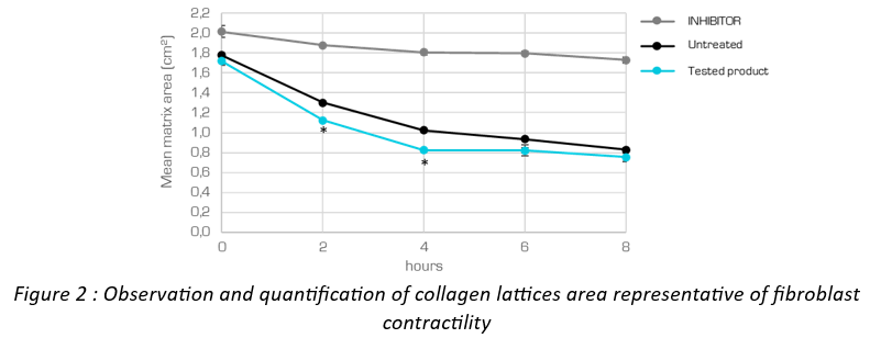
By exerting traction on the matrix, fibroblasts can influence its reorganization and density. This leads to a reduction in the surface area of the gel. The amount of retraction (gel surface) can be measured as an indicator of fibroblast contractility (Figure 2).


Quantification of stress fibers
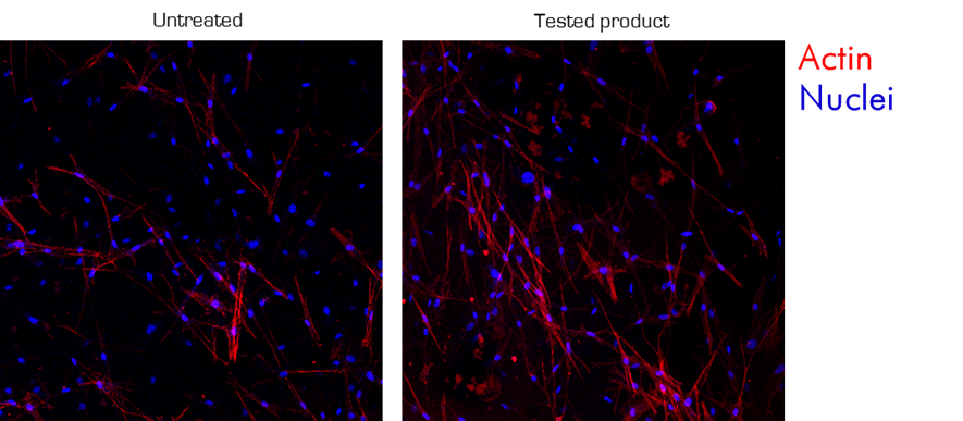
Actin is a globular protein which, when polymerized, forms actin filaments, the main components of the cellular cytoskeleton. These actin filaments are crucial for many cellular processes, including motility, division and cell contractility. In fibroblasts, actin filaments are organized into structures called stress fibers. Fibroblast contractility is mediated by interactions between actin filaments and myosin within stress fibers.
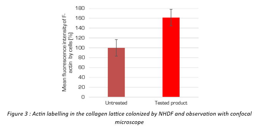
It can study the quantity and organization of fibroblast stress fibers within each collagen gel. (figure 3).


The number of stress fibers within dermal fibroblasts, and their organization, can reveal several important aspects of the cell’s physiological state. An increase or reorganization of stress fibers globally indicates cell activation for wound healing, extracellular matrix neosynthesis or cytoskeletal reinforcement.
TO IMPROVE HEALING AND CONTERACT AGING
In conclusion, this in vitro model provides a means of studying skin fibroblast contractility and understanding how it is regulated.
This type of study, and further investigation of this mechanism, will lead to a better understanding of how fibroblast contractility influences skin healing and aging, as well as the effects of ingredients on these processes.
*publirédactionnel
CONTACT
Claire Leduc


 Follow us on Linkedin!
You must be logged in to post a comment.