Promega’s 3D cell-based assays: Modern tools to monitor skin biology in 3D culture
2 December 2024
The cosmetics industry is undergoing a profound transformation, driven by advances in scientific methodologies and heightened regulatory and consumer demand for ethical and reliable testing. Among the most groundbreaking changes is the shift towards in vitro 3D models.
From 2D to 3D: A Paradigm Shift
While 2D cultures provide valuable insights, they fail to accurately mimic the complexity of human skin. Cells grown on flat surfaces lack structural depth and intricate interactions present in real tissue, which can lead to discrepancies between lab results and real outcomes.
3D cultures systems better mimic the three-dimensional architecture tissue more effectively than monolayer cultures, enabling more realistic cell-cell and cell-extracellular matrix (ECM) interactions that influence cell behavior. This makes 3D systems, ranging from simple spheroids to complex microphysiological systems with interconnected organoids on microfluidic chips, more physiologically relevant models for studying skin biology.
3D Cell Models: With Great Promises Come Significant Challenges
The use of assays originally designed for cell monolayers or suspensions may yield inaccurate or misleading results when used with 3D models. For instance, MTT and resazurin assays are not optimized for 3D systems. Their poor reagent diffusion often underestimates cell viability, and prolonged incubation times—necessary for adequate penetration into 3D structures—can lead to cytotoxic effects, as both MTT and resazurin reagents have been observed to be toxic to cells. Additionally, MTT is known for its low sensitivity and potential interference with ECM components, further limiting its compatibility with 3D cultures
Promega’s 3D-Validated Cell Health Assays
The primary question after treating cultured cells, including in 3D models, is whether the treatment affected cell viability. This is assessed using either a cell viability assay, which measures live cells with intact membranes, or a cytotoxicity assay, which measures dead cells with compromised membranes.
Many Promega assays have already been validated to show good performance and produce accurate results with different 3D cell culture models. Others were specifically optimized with adapted reagents.
In 3D models, ECM coatings often hinder reagent penetration, leading to incomplete lysis and inaccurate results, as seen with the original CellTiter-Glo® Cell Viability Assay. To address this, the assay was reformulated into CellTiter-Glo® 3D by enhancing its lytic capacity with additional detergents and longer exposure times, enabling deeper penetration and complete lysis of spheroids.

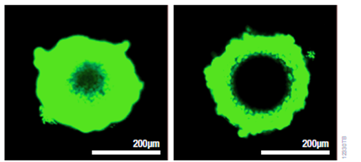
Improved 3D microtissue penetration and more accurate viability data HCT116 colon cancer spheroids were generated by seeding cells in the InSphero GravityPLUS™ 96-well hanging-drop platform and grown for 4 days. A 2X concentration of CellTox™ Green Dye was added to CellTiter-Glo® 3D Reagent (left) or other reagent (right) prior to sample addition as an indicator of cell lysis and images were acquired at 30 minutes. The spheroids in Panel B are ~300μm in diameter, and the bars in each image represent a distance of 200μm.
Some assays do not require modification; for example, the RealTime-Glo™ MT Cell Viability Assay, LDH-Glo™ Cytotoxicity Assay, and CellTox™ Green Cytotoxicity Assay work effectively with many 3D models without changes.
Promega offers many other cell-based assays tested with 3D cultures for cell viability, cytotoxicity, apoptosis, metabolism, inflammation and more, to help researchers get reliable tools for studying 3D models.
“All Models Are Wrong, But Some Are Useful”, And Cruelty Free
Promega’s 3D cell-based assays leverage sophisticated 3D models to deliver superior insights into product safety and efficacy, without reliance on animal testing.
To simplify the adoption of these technologies, Promega offers technical and scientific support, including detailed protocols, training sessions, and a dedicated applicative support service.
CONTACT
Promega France


 Follow us on Linkedin!
You must be logged in to post a comment.