
A Validated Method for Assessing the Effects of Blue Light on the Human Skin by Genemarkers
1 October 2023
Rishabh Kala, PhD*, Nicole Heiberger, MS, Heather Mallin, BS, Stephanie Wheeler, MS, Anna Langerveld, PhD
To read the article published in the International Journal of Cosmetic Science: click here
Introduction
The COVID pandemic caused an increase in virtual meetings and work from home scenarios that resulted in people spending increased time indoors and in front of computer screens and electronic devices. It is estimated that 60% of people spend more than six hours per day in front of a digital device.
Studies have shown that blue light, also known as high-energy visible (HEV) light, can produce cytotoxic effects, primarily through the production of reactive oxygen species and increased inflammation. However, the topic has been controversial with some studies claiming no adverse effects of blue light on the skin.
Methods for testing the effects of blue light using in vitro testing models are lacking. This work was conducted in order to develop a reproducible, validated testing method for assessing the effects of blue light on the human skin.
Methods
We designed a custom blue-light box that generates blue light at 460 nm that can be maintained in an incubator to support in vitro testing. We performed a series of studies to optimize the dose and timing of the exposure, cell culture conditions, and the extraction of nucleic acid and protein for gene and protein analysis. Immunohistochemistry (IHC) for select proteins was performed to visualize changes in the protein levels in the different cell types and/or layers of the skin tissues.

Tissue Model:
The studies were performed using full-thickness EFT-400 in-vitro skin tissues comprised of normal human keratinocytes in a stratified corneal layer, and a dermal component containing an intact
dermal-epidermal junction (DEJ) and viable fibroblasts. The cells that are utilized to produce the tissues are derived from neonatal tissue sources. Tissues were cultured according to the manufacturer’s recommendations (37°C, 5% CO2) using antioxidant and phenol-red free media. Tissues were exposed for either 1, 2, or 6 hrs for 5 consecutive days. The intensity of the blue light exposure was 30 J/cm2. Studies were repeated 3-4 times to assess reproducibility.
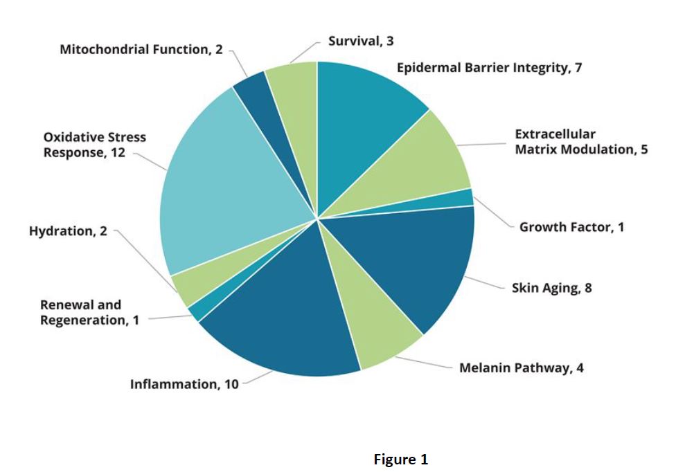
Gene Expression:
Gene expression was assessed using Genemarkers’ Standard Skin, Environmental Stress, and Brightening Panels, which contain TaqMan® qPCR assays formatted into arrays, assessing
a total of 290 target genes and 5 endogenous control genes.
Statistics were performed using ThermoFisher Connect RQ software, with gene expression levels compared relative to unexposed
control tissues.
Protein Expression:
Enzyme-linked immunosorbent assays (ELISAs) for GM-CSF, 8OH-dG, and MMP1 were performed according to the manufacturer’s instructions. Data was analyzed relative to a standard
curve. Statistical analysis was performed using a Student’s t-test (unpaired; p< 0.05).
Protein expression

Exposure to blue light significantly increased 8-hydroxy-2′-deoxyguanosine (8-OHdG), a marker for oxidative stress, and MMP1 and CSF2, markers
for aging and inflammation (Fig.3).
Immunohistochemistry (IHC) was performed for collagen, filaggrin, and NAD(P)H quinone dehydrogenase 1 (Fig. 4) to correlate the changes in gene expression with protein changes and to visualize the effects in different layers of the skin tissues. Exposure to blue light produced changes in the amount and localization of collagen, filaggrin, and NQO1.
gene expression
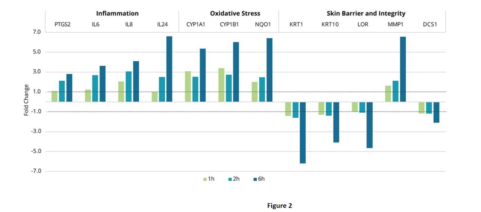
Conclusions
► This work demonstrates a reproducible in vitro testing method for assessing the effects of blue light on the skin.
► The data shows that blue light produces negative effects on the skin via increased inflammation and biomarkers for the response to oxidative stress. In addition, exposure to
blue light decreased expression of important genes that regulate epidermal barrier function and tissue integrity.
► This work validates the use of Genemarkers’ new Blue Light testing service.
Publi redactionnel – Sponsored ‘advertorial
Contact
Tel: +1 269-808-1517
email: amorrison@genemarkersllc.com
Web site: https://genemarkersllc.com
126 E. South Street
Kalamazoo MI 49007 – USA
![]()


 Follow us on Linkedin!
You must be logged in to post a comment.