Skin analysis can be performed by various measurements on the skin surface in combination with a visual assessment by eye, or by using a dermatoscope. To get additional information about the composition of the skin layers, non-invasive real-time ultrasound imaging can be used as a highly recognised and widely used scientific research method. The deeper insight is useful when assessing the effect of topical treatment, collagen supplements, laser therapy, or subcutaneous fat reduction.
How does ultrasound skin imaging work?
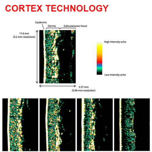
An ultrasound image is a visualisation of the acoustic properties of the tissue under investigation. Short acoustic pulses are transmitted into the skin and reflections from the pulse are recorded using an ultrasound transducer. The energy of the acoustic pulse is very low and does not cause any adverse effects such as heating or cavitation. Special software is used to analyse the ultrasound signals and calculate the intensity (energy) of the reflected signals in specific skin layers. In specific skin layers, the intensity has been shown to correlate well with the amount of collagen. Other parameters such as skin thickness and age-bands (LEB) can be determined automatically by the software (see below image on age differences (Fig. 2)).
What can you see with ultrasound images?
The ultrasound image is a cross-sectional view of the tissue where each pixel has a colour based on the signal energy (Fig. 1). The more homogeneous, the darker the colour (e.g., fat or water). A brighter colour represents structures with more reflections due to acoustic impedance, non-homogeneous composition, and density differences. evalau
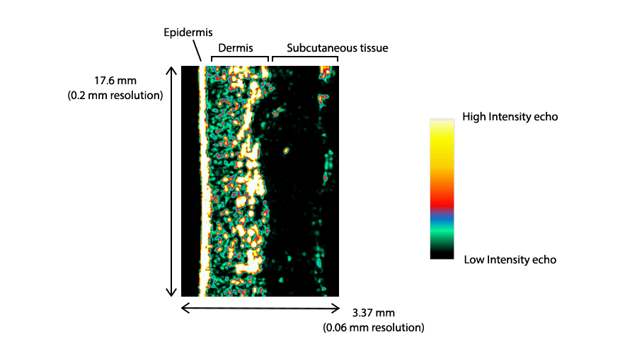
Figure 1. A cross-sectional view of the tissue where each pixel has a colour based on the signal energy.
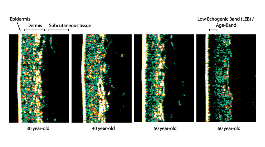
Figure 2. The collagen represented in the picture as bright colors decrease with age.
Skin ultrasound technology for every research application
Cortex Technology, the leading provider within skin ultrasound technology, offers a selection of different ultrasound probes depending on the application.
DermaScan C ultrasound linear scanning system consists of 3 different probes with 20-50 MHz transducers individually adapted to the actual probe, providing scanning depts of 7-23 mm penetration. 6-8 frames per sec with automatic cineloop. A dynamic scanner used in a broad range of product research & development, and clinical research studies. References can be provided upon request.
DermaLab Combo SkinLab besides a wide range of analysing probes also features unique ultrasound probes.
Standard high frequency ultrasound probe: a rotating single element transducer with a centre frequency of 20 MHz giving a still scan with a dept of 3.4 mm. The B-mode scanning makes skin thickness and density measurement easy and instant. Resulting in Collagen level assessment, dermal thickness reading and identification of the lower echogenic band (LEB)/ Age-band. References can be provided upon request.
Subcutaneous ultrasound probe: a rotating single element transducer with a centre frequency of 10 MHz giving a still scan with a dept of 40 mm. As the name indicates, this special probe is designed to be used to visualise the structures of the subcutaneous layers. For example, it is seen to be useful in non-invasive subcutaneous fat reduction treatment to evaluate the treatment success. References can be provided upon request.
For further information: https://cortex.dk/skin-analysis/
Join us: https://www.linkedin.com/company/cortex-technology-aps/mycompany/
www.cortex.dk | Corporate and testing sheet: https://skinobs.com/instrumentation.php?id=90
Read the entire ZOOM#23 here
CONTACT
![]()


 Follow us on Linkedin!
You must be logged in to post a comment.