Microscopic examinations and analyses are considered the gold standard for assessing pathogenic tissue changes. Invasiveness, a restriction of the diagnostics to the area from which the tissue sample was excised, and a time delay between sample extraction and providing a diagnosis are disadvantages of traditional methods.
In vivo examinations using confocal laser scanning microscopy have the benefit of a non-invasive procedure and time savings. This method facilitates the precise differentiation by the physician between pathogenic and healthy tissue and is not restricted to an excised tissue sample. The examined skin regions remain unchanged and are available for subsequent standard histological methods.
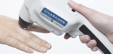
The in vivo devices VivaScope® 1500, VivaScope® 3000, and VivaScope® 1500 Multilaser deliver an in-depth view into living tissue within its natural environment – confocal laser scanning microscopy opens a window into skin. Reflections caused by differences in the refractive index of the constituents of skin make it possible to use confocal laser scanning microscopy without external contrasting agents.
MAVIG offers confocal laser scanning microscopes for in vivo and ex vivo use. All units are shipped with specially developed software. A handheld unit was developed as an especially flexible device. As a supplement to the confocal in vivo images, a digital macro camera can take images of skin to ensure optimal examination conditions.
MAVIG also offers a comprehensive service for all of its VivaScope® devices. After the devices are installed on-site, service includes connection to the local network, introduction and training with the device, as well as advanced expert training.


 Follow us on Linkedin!