Study of the Sensitive Skin and the “Anti-inflammatory” Effects by Miravex
12 June 2023
Sensitive skin is a common and frequently occurring skin disorder that may affect about 30–50% of the global population, which can be manifested as facial erythema accompanied by pruritus, burning or tingling sensation, and skin tightness.
The measurement of haemoglobin content and its distribution are important parameters to evaluate skin inflammation, and the Antera 3D is particularly well suited to perform these measurements.
Unlike traditional imaging techniques, where only three colour channels (red, green, and blue) are used, the Antera 3D® uses reflectance mapping of seven different light wavelengths spanning the entire visible spectrum. This allows for a more precise analysis of the skin colorimetric properties, which are mostly determined by two dominant chromophores: melanin and haemoglobin.
Acquired spectral images are transformed into skin spectral reflectance maps, and the skin surface shape is used to compensate for light intensity variation due to the varying direction of incident illumination. The reflectance data is transformed into skin absorption coefficients and used to quantify melanin and haemoglobin concentrations using mathematical correlation with known spectral absorption data of these chromophores[1]. The acquired spectral data is used to map the distribution and concentration of melanin and haemoglobin, providing a visual representation of their concentration and distribution in the skin. The result is repeatable measurements of spatially resolved haemoglobin/melanin.
The Antera 3D has been used extensively in many publications to measure vascular lesions, skin sensitivity and inflammation – a full bibliography is available at https://miravex.com/publications/. Here, we will briefly discuss a recent publication[2] that has proposed the Antera 3D as a method to objectively detect and quantitatively evaluate sensitive skin by analyzing texture, hemoglobin concentration and the area of the vascular lesions.
[1] R.R. Anderson and J.A. Parrish. “The optics of human skin”. Journal of Investigative Dermatology, 77 (1981), pp.13–19
[2] X Ai’e et al., “Quantitative evaluation of sensitive skin by ANTERA 3D® combined with GPSkin Barrier®”, Skin Res Technol. 2022;28:840–845.
In this study, 20 subjects with sensitive skin were treated with an anti-sensitive moisturizing tolerance-extreme cream which has anti-inflammatory and moisturizing effects, twice daily on the whole face for 28 days. Texture, haemoglobin, and influenced area (mm2) were recorded using ANTERA 3D. Subjects underwent skin tests and recorded changes at D0 and D28. 20 healthy participants were recruited and used as control.
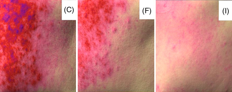
Compared with controls, the hemoglobin concentration and influenced area of sensitive skin subjects – measured with ANTERA 3D – were significantly higher than healthy participants’ data. Compared to D0, both a clear trend toward decreased hemoglobin and influenced area of sensitive skin subjects were seen at D28. It was also observed that the skin texture improved significantly at D28.

Figure 1. ANTERA 3D images of sensitive skin subject at D0 (C), sensitive skin subject at D28 (F) and control subject (I).
In conclusion, thanks to its repeatability, high specificity[1] (i.e. the ability to effectively separate the melanin and haemoglobin components from the reflectance spectra), and spatially resolved spectral response, the ANTERA 3D can be used to detect and quantitatively evaluate sensitive skin.
[1] Matial et al., “Skin colour, skin redness and melanin biometric measurements: comparison study between Antera” 3D, Mexameter” and Colorimeter”, Skin Research and Technology 2015; 0: 1–17.
CONTACT
+353 (0)1 525 2865


 Follow us on Linkedin!
You must be logged in to post a comment.