Why has the most universally sought-after claim in cosmetics never ceased to reinvent the way in which it can be scientifically substantiated?
Hydration remains the most systematically supported claim in clinical studies, regardless of product category, country, or consumer target. Brands developing anti-ageing, soothing, radiance-enhancing, anti-pollution or barrier-repairing formulas all ultimately face the same fundamental question: how does this product act on skin hydration, and how can it be scientifically proven?
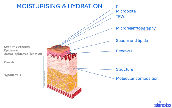
It is the proof itself, its nature and its scientific component, that concentrates all the complexity of evaluating this product’s performance. Skin hydration is not a surface phenomenon occurring in isolation. It traverses all layers, stratum corneum, epidermis, dermis, from the surface to the dermal matrix, and contributes to the overall equilibrium of the skin. Essential to life, water homeostasis maintains the water gradient from depth to surface and contributes to barrier function. It is interdependent with various essential mechanisms such as epidermal renewal, pH regulation, temperature regulation, microbiome balance, immune and inflammatory equilibrium, and hormonal and neuroendocrine homeostasis. The importance of the skin’s response to environmental stress and the overall impact of the exposome must also be considered. Measuring “hydration” in a meaningful way requires deliberately choosing which layer and which mechanism one intends to capture.
Towards a Multi-Modal Approach
The modern beauty routine is perceived in an integrative manner, encompassing quantified performances, emotional balance, and lifestyle. Within this holistic vision, hydration is both a visible indicator of skin condition and balance, and a symbolic expression of vitality. Consumers seek measurable structural improvements, neurosensory pleasure, and enhanced self-esteem.
In in vivo volunteer studies, whether clinical protocols with expert evaluation or biometric measurements, descriptors such as “hydrated” must be translated into intelligible, meaningful, and reproducible outcomes. The in vivo assessment of hydrating effects relies on the use of various methodologies:
- Consumer tests with self-assessment: these tests reflect the users’ perception of the product. Participants indicate their perception of hydration using structured questionnaires and rating scales. These tests capture the subjective improvement expected by consumers and are essential for substantiating claims.
- Clinical evaluation by scoring: semi-quantitative assessment carried out by qualified evaluators. Trained dermatologists or cosmetologists assess hydration using validated scales under standardised lighting and imaging conditions. This method reduces, though does not eliminate, subjectivity.
- Sensory analyses and neurosensory measurements: these capture unconscious or emotional responses. The study of emotional processes represents an inexhaustible source of innovation for the cosmetics industry. It is essential to combine the study of the three emotional components: behavioural, physiological, and cognitive, and to pay particular attention to methodological rigour. Emerging methods such as facial expression analysis, eye-tracking, and EEG measure unconscious reactions.
- In vitro/ex vivo assays and specific biomarker analysis.
- In vivo biometrological measurements: objective and instrumental measurements of the optical properties of the skin. A broad range of optical and imaging devices enables the objective quantification of hydration.
The Reference: Instrumental Dielectric Measurement
Since the 1980s, the reference approach has been based on the electrical behaviour of the skin. The Corneometer was the first device to be commercialised by the German company Courage & Khazaka and has become the universal standard for the evaluation of skin hydration. Today, the Corneometer CM 825 measures capacitance over the first 10 to 20 µm of the stratum corneum and serves as the benchmark; other hydration measurement instruments being calibrated by comparison with it.
Currently, a large number of probes (17 referenced to date on the Skinobs platform) measure capacitance, impedance or permittivity to quantify the water content of the skin, with devices adapted to each depth:
- The stratum corneum: Corneometer CM825, Dermalab, Epsilon, MoistureMeter SC, Skicon-200, DPM 9003, EveKey Skin
- The epidermis: MoistureMeterEpiD, Nevisense
- The dermis: MoistureMeterD
New devices based on this same technology are regularly emerging, such as the Nevisense (Scibase), Z-Pen (Clarins-Piwio), and particularly for novel nomadic applications intended for home diagnostic devices. The value of these devices is as much operational as scientific: rapid, reproducible, usable in all climatic conditions and across all skin ethnicities, and well understood by regulators, testing laboratories, cosmetics brands, and ingredient manufacturers alike.
Alternative Measurements of Interest
The measurement of transepidermal water loss (TEWL) takes an indirect route by monitoring the moisture gradient escaping from the skin surface. Particularly useful for measuring long-term effects and lipid-rich formulas, TEWL serves as a general marker of barrier function. Indeed, barrier integrity will determine whether hydration is maintained over time, and not merely at the moment of application. The devices used (6 referenced on the platform and used by more than 100 CROs): Tewameter 330, Tewameter HEX, Tewameter Nano, DermaLab, Aquaflux, Vapometer, RG1 Evaporimeter.
The skin microbiota and skin hydration maintain a close interdependent relationship. A well-hydrated stratum corneum provides a favorable environment for commensal bacteria, notably Staphylococcus epidermidis, which contributes to maintaining the acidic pH of the cutaneous mantle. This acidic pH in turn governs the activity of lipid enzymes involved in the synthesis of the intercorneocyte cement, thereby reinforcing the barrier and limiting transepidermal water loss. Conversely, dysbiosis impairs barrier integrity and promotes skin dryness. Jointly assessing microbiota and hydration through in vivo collection methods (swabbing/stripping/curling, etc.) coupled with multi-omic analyses opens new perspectives for objectively demonstrating efficacy in dermato-cosmetics.
Furthermore, a broader palette of tools has been developed, quantifying and/or visualising directly the quantity of water, lipids, and their impact on skin structure through optical and spectroscopic methods:
- In vivo microtopography (MoistureMap 100-200, Epsilon E100), with a conversion of conductivity measurements into a greyscale image
- Microtopography from stripping: XFluo-3D Fluorescence technology or Atomic Force Microscopy
- Near-infrared spectroscopy, NIRS, Dermo
- Confocal Raman spectroscopy: Raman spectroscopy gen2-SCA, LabRam 800, Aqualog
- Opto-thermal emission radiometry (OTTER)
- 2D surface imaging: Visioscan VC, C-Cube, EvaSurf, Dermalab, SkinCam, DigiCam, SpectraFace, Derma Reader/Scope, LifeViz, Hirox, VideometerLab, etc.
- 3D surface imaging: Antera 3D, Visia 3D
- Study of lipids, their quantity, distribution, and composition to better assess skin hydration equilibrium phenomena, via HPLC measurements, transmission electron microscopy, etc.
High-Resolution and Molecular Readings
The frontier of measurement has shifted towards non-invasive techniques that directly visualise skin structure rather than inferring it from surface signals. Multiphoton tomography, confocal microscopy, and Raman spectroscopy now make it possible to observe water distribution and molecular composition in situ, layer by layer.
Among these, LC-OCT (Line-field Confocal Optical Coherence Tomography) merits particular attention. Initially developed for dermatological diagnosis (detection of skin cancers, characterisation of lesions), LC-OCT delivers 3D imaging combining quantitative depth data with a genuinely visual, almost histological, non-invasive and biopsy-free reading of the skin. What is now emerging is its extension towards the substantiation of cosmetic claims: a diagnostic-grade instrument finding a second application by associating quantified hydration data with visual proof, complementing the numerical result expected by both regulators and marketing teams. This is a compelling example of how clinical evaluation is no longer limited to producing a figure: it now produces a figure that can be visualised.
The Mechanistic In Vitro Approach
All this instrumental evidence means little without an understanding of what occurs at the cellular scale; this is where in vitro and ex vivo work finds its place, not as a substitute for the clinical study, but as a mechanistic explanation.
Hyaluronic acid remains the reference molecule, prized for its almost unrivalled capacity to bind water within the extracellular matrix. The natural moisturising factor (NMF), those free amino acids released by the degradation of filaggrin, governs flexibility, desquamation, and the overall homeostasis of the barrier at the level of the stratum corneum. Aquaporins, those membrane proteins forming water channels, decline with age, a phenomenon increasingly recognized as a direct driver of progressive skin dehydration; this also explains why anti-ageing actives that support aquaporin expression carry a hydrating benefit almost by mechanism, and not merely by association.
Other markers merit monitoring: glycosaminoglycans and proteoglycans, which help retain water within the dermal matrix; CD44, the principal membrane receptor for hyaluronic acid; caspase-14, which drives the proteolysis of filaggrin into NMF precursors; ceramides and phospholipids, whose physical organisation within the lipid bilayers of the stratum corneum is itself a determinant of hydration status; and matrix metalloproteinases, involved in the remodeling that either sustains or weakens the density of the extracellular matrix over time.
These markers can be monitored on several type of assay supports 2D cell lines, co-cultures, iPS-derived models, 3D organoids, or complete reconstructed skin models, bioprinted or otherwise, by gene expression, histology, or protein assays (ELISA, Western Blot). Dehydration itself can be simulated in vitro, typically by increasing salt concentration or withdrawing culture medium, enabling protective or reparative ingredients to be tested well before the design of a full clinical study.
Designing the Right Protocol
Hydration claims can be substantiated across very different time horizons: short-term effects between 30 minutes and one hour, intermediate effects over one day, or sustained effects over one to four weeks. The choice of device, panel size and inclusion criteria, and acclimatization protocol depends entirely on the objectives defined by the product claims. A formula rich in electrolytes or small humectant molecules may artificially inflate impedance-based readings, just as a lipid-rich formula may underestimate them; these are not flaws in the method, but reasons to select the appropriate combination of methods suited to the formula. This is precisely where early and detailed exchanges with testing laboratories prove their worth, by defining inclusion criteria, measurement schedule, treatment conditions, and instrumentation before the protocol is finalized.
The Next Step: Combining Biomarkers and AI
The truly prospective development is not a new standalone device; it is what occurs when this molecular evidence is combined with artificial intelligence. Rather than reading biomarker panels, images, and biophysical measurements as separate, siloed datasets, AI-driven analysis can cross-reference them, revealing correlations that would remain invisible to a researcher examining each technique in isolation. For hydration in particular, this means models trained to link, for instance, aquaporin or filaggrin expression to TEWL trajectories and structural changes observed in imaging, with the possibility of predicting which formulations will perform clinically before even launching a full in vivo study. The practice remains emergent rather than standardized, but it signals the direction in which claims substantiation is heading: a multimodal approach combining biological mechanisms, biophysical measurements, and visual evidence, all interpreted in light of all the data generated.
Key Takeaways
Hydration testing has not become simpler as the palette of available tools has expanded; if anything, the reverse is true. Yet these technological advances are good news for brands and ingredient manufacturers: the current combination of instrumented biophysics, imaging techniques such as LC-OCT, compositional techniques such as Raman spectroscopy, and the nascent coupling of molecular biomarkers with AI-driven analysis now offers the means to substantiate a hydration claim with a scientific depth and credibility that simply did not exist ten years ago. The brands that make the best use of this field of investigation are not necessarily those that multiply tests; they may rather be those that choose the right combination, conceived from the outset in relation to their primary and secondary claims.
In conclusion, hydration is defined by a multidimensional synergy of biological, optical, and perceptual determinants. Its validation requires an integrated framework combining the study of in vitro mechanisms, subjective consumer feedback, expert evaluation, and advanced biometrology. Modern assessment favors 2D/3D imaging, non-invasive microscopy, and measurements of the dielectric component of the skin. Ensuring data integrity demands standardized protocols focused on precise spatial repositioning and robust acquisition (technician/machine). The future of this performance lies in the convergence of high-definition optical sensors and AI algorithms, which bridge the gap between neurosensory perception and physiological parameters.
| Type of methods | Studied effect | Rôle | Depth in the skin | Imaging | Imaging & quantification | Quantification |
|---|---|---|---|---|---|---|
| Hydration evaluation | Dielectric component of the Stratum Corneum | Evaluation of the Hydration content via the relation of the hydration rate and the response of the dielectric component of the tissue through the capacitance, permitivity… | Stratum corneum | Epidermis | Epsilon (Biox), Skicon-200 | Corneometer (C+K), DPM 9003 (NovaTech), Moisturemeter SC/D/epiD (Delfin), Dermo (Varennes), Epsilon (Biox), DermaLab (Cortex); Nevisense (Scibase), Skicon-200 (IBS-Japan) | |
| Optical by Infra-red | Evaluation of the Hydration by reflexion of the Near Infra-red closed to the hydration transmission bands | Stratum corneum | NIR Spectroscopy | |||
| Spatial distribution of the hydration & microtopography of the skin surface | Evaluation of the Hydration content via the response of the capacitance. | Stratum corneum | MoistureMap (C+K), SkinChip (L’Oréal) | |||
| Hydration gradient | Optical technologies via Laser excitation and thermal analyse | Stratum corneum | Epidermis | Opto-thermal emission radiometry (OTTER) | |||
| Composition of the skin | Biomarkers of the Stratum Corneum | General biomarkers (NMF, Filagrine…) involved in the hydration balance after skin strippings | Stratum corneum | SquameScan (Heiland), Genomic, metabolomic, proteomic, multi-omics, HPLC… | ||
| Collagen content | The water contained in the collagen network gives the dermis its viscoelastic properties. It maintains, with the GAGs, an interstitial pressure gradient that directs the flow of water towards the upper layers of the epidermis. | Dermis | Scanning electron microscopy (SEM) | SIAScope (Medxhealth) – Dermo (Varennes) | ||
| Molecular content & distribution in the skin | For specific molecules, keratin, collagen, lipids, water, hyaluronic acid involved in the Hydration balance | Stratum corneum | Epidermis | Dermis | LC-OCT (Damae) | Aqualog, Sonde Raman (Horiba), Raman spectroscopy gen2-SCA (RiverD), Genomic, metabolomic, proteomic, multi-omics… | ||
| Sensory & perception | Softness & tribology of the skin surface | Stratum corneum | Frictiometer (C+K), Touchy Finger Tribologie (Tactinnov) | |||
| Surface of the skin | Nanotopography of the corneocytes via stripping | The surface, the roughness and the shape of the keratinocytes give information of the quality of the Stratum corneum. | Stratum corneum | Xfluo (Kamax), ECTI, CNOs (Loretta) | ||
| Surface visualization and Microrelief network | General evaluation of the skin conditions | Stratum Corneum | Videomicroscope (Hirox), Dermascope (Dino-lite), DermLite DL100, Videometer Lab, VEOS DS3 (Canfield), DermaLab Videoscope (Cortex) | Antera 3D (Miravex), DermaTOP-HE-60, EvaSurf (Eotech), SpectraCam, SkinCam (Qima Life Sciences), Visioscan (C+K), Visia CR (Canfield), TiVi 60 Skin Damage Visualizer (Wheelsbridge), SIAScope (Medxhealth), DermaTOP-HE-60 (Eotech), C-Cube (Pixience) | ||
| Surface caracterisation | General evaluation of the skin conditions | Stratum Corneum | Scorage, sensory panel trained or naive panel, neuro-psychosensorial (EEG, Mood Board, Quality of Life, f-IRM…) | |||
| Structural mechanisms | Epidermis renewal | Keratinocyte turnover is a major determinant of skin hydration because it conditions both the quality of the stratum corneum, the effectiveness of NMF and the integrity of the lipid barrier. | Epidermis | DHA or Dansyl Chloride marking and Skin color analysis | ||
| Lipids fractions & distribution | The main function of skin lipids (ceramides, cholesterol) in hydration is a barrier function that directly conditions water retention in the intercorneocytar space and all skin layers. | Stratum corneum | Transmission electron microscopy (TEM), Immuno-fluorescence Microscopy, Fluorescence Light microscopy, HPTLC | Fourier transform infrared spectroscopy (FTIR ATR), Chromatography (HPTLC, LC/MS), OTTER | ||
| Structure: visualization by Microscopy | Direct and non-invasive 3D observation of the skin structure, the different cell layers and the cells. | Stratum corneum | Epidermis | Dermis | Vivascope (Mavig), Vivosight (Michelson) | LC-OCT (Damae) | / | |
| Structure: visualization by Ultrasound | Direct and non-invasive 2D observation of the skin structure, the different cell layers and the cells. | Stratum corneum | Epidermis | Dermis | Dermascan, Dermcup, Dermalab (Cortex), DUB®SkinScanner 50/22 (Eotech), Ultrasound WED-2018 (Wed) | / | ||
| Transepidermal Water Loss by TEWL | The water evaporation of the skin evaluates the skin barrier’s ability to retain skin water and maintain the hydration gradient | Stratum Corneum | Aquaflux (Biox), Vapometer (Delfin), RG1 Evaporimeter (Cyberderm), Tewameter 330, Tewameter HEX, Tewameter Nano (C+K), Dermalab-TEWL (Cortex) |