Hydration is one of the most common claims in the cosmetics industry. Whether it’s facial, body or scalp care, demonstrating the moisturizing effectiveness of a product is an essential step in its development.
To do this, laboratories now have robust and widely recognized instrumental methods. Corneometry is used to assess the water content of the surface layers of the skin, while the measurement of insensible water loss (TEWL) provides information on the integrity of the skin barrier function.
These techniques provide much-needed quantitative data, but they don’t always describe the visible consequences of hydration. However, better hydrated skin generally appears less dry, smoother, more homogeneous and more radiant.
It is precisely these visible effects that contribute to consumers ‘ perception of the product’s effectiveness . It is in this context that clinical imaging takes on its full value.

What the C-Cube brings to hydration assessment
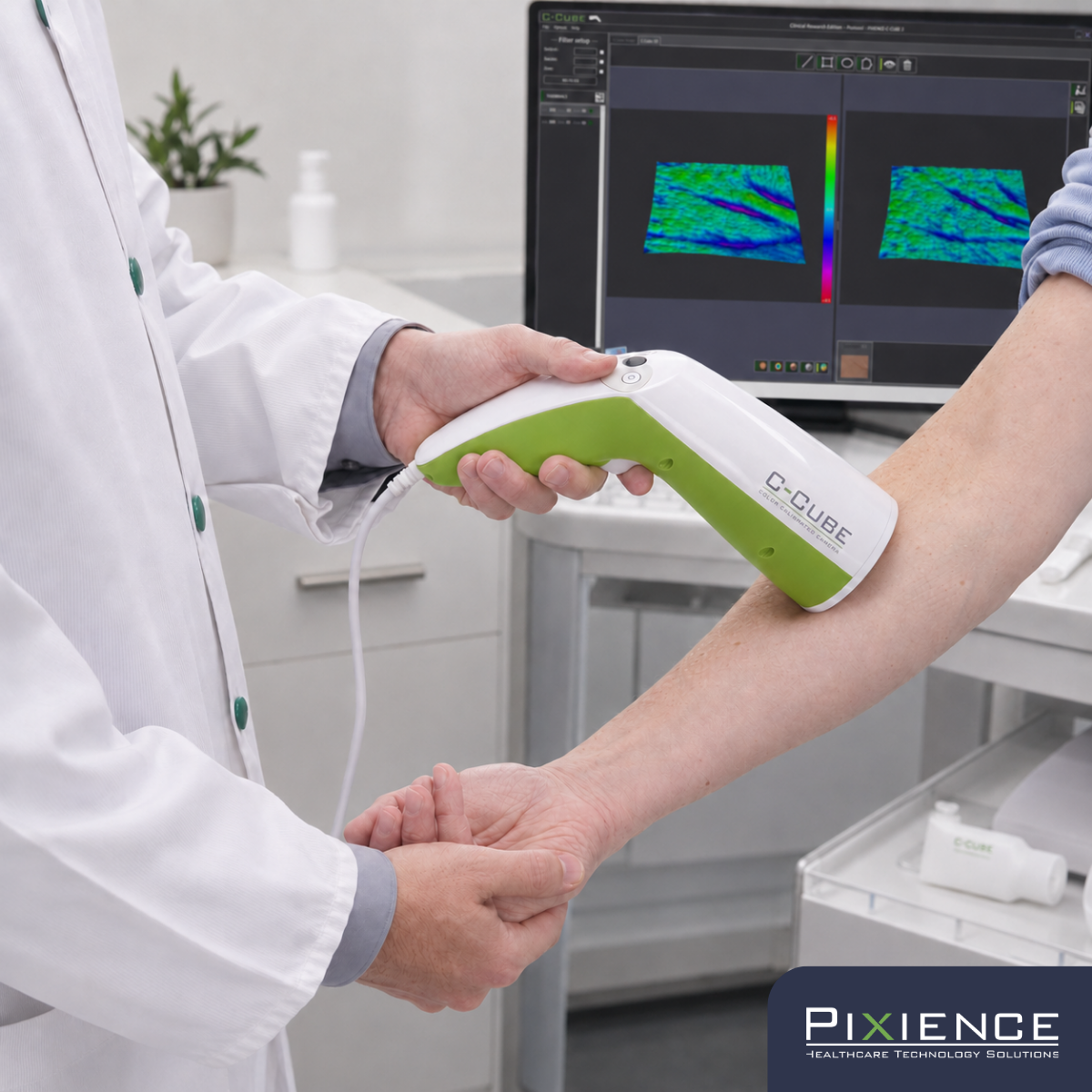
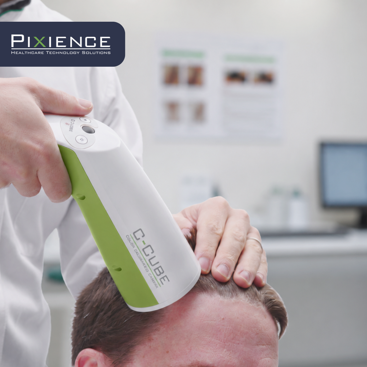
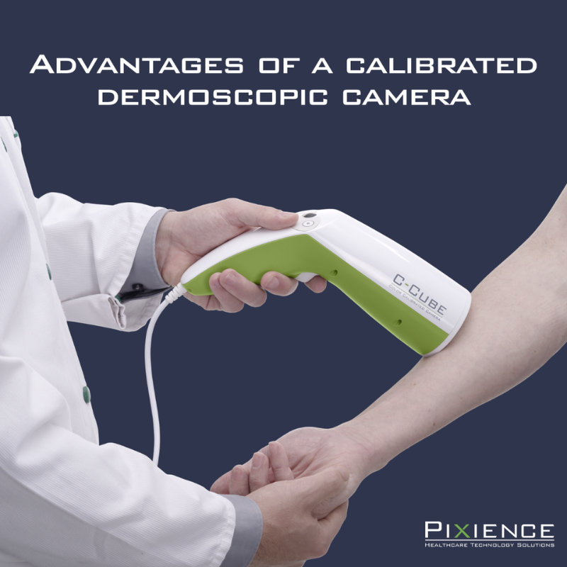
The C-Cube Clinical Research was designed to transform visual observations into objective, quantifiable data.
Its technology is based on three complementary pillars:
- High-resolution microdermoscopic imaging, allowing the visible signs of skin dryness to be closely observed;
- Colorimetric and metric calibration, guaranteeing the reproducibility of measurements over time;
- A three-dimensional reconstruction of the skin surface, making it possible to quantify the texture and organization of the skin’s relief.
This combination makes it possible to objectively visualize and quantify the visible consequences of hydration in clinical studies.
From image to measurement
One of the main strengths of the C-Cube is its ability to turn an image into a measurement.
The 3D reconstruction of the skin surface makes it possible to calculate various roughness and texture parameters, such as Sa, Sq or Sdr, which are widely used in the analysis of surface conditions.
The system also makes it possible to evaluate the organization of the skin microrelief thanks to parameters such as isotropy, which characterizes the distribution of furrows on the surface of the skin.
Well-hydrated skin generally has a more homogeneous and better organized microrelief than dry or altered skin.
Thus, beyond the visual aspect, the observed changes can be quantified and statistically monitored throughout the study.
Why standardization is essential
The use of imaging in a clinical context imposes a fundamental requirement: reproducibility.
Without careful control of acquisition conditions, variations in lighting, color, or magnification can mask the true effects of a product. The C-Cube incorporates colorimetric and metric calibration ensuring that the observed differences reflect the effects of the product and not variations in acquisition.

Combining biological data and visual evidence
Let’s take the example of a study conducted on subjects with dry to very dry skin. After several days of using a moisturizer, corneometry can show a significant increase in the water content of the stratum corneum.
Imaging then provides additional information by showing how this improvement becomes visible on the surface of the skin:
- Decreased scales;
- Reduction of signs of xerosis;
- Improved roughness;
- Homogenization of the microrelief;
- Overall improvement of the appearance of the skin.
Instrumental measurements explain what is happening in the skin; Imagery makes it possible to objectively document what becomes visible on its surface.
The Growing Importance of Visual Evidence
The results of a clinical study are no longer only intended for the R&D teams. They must also be able to be understood and valued by marketing teams, brands and consumers.
In this context, standardized images, analysis maps and 3D reconstructions are particularly powerful tools. They illustrate the benefits observed while maintaining a high level of scientific rigour.
The parameters resulting from the imaging are of particular interest because they correspond directly to the benefits sought by consumers: softer, smoother, less dry, more homogeneous and visibly healthier skin.

Join us to explore these approaches in detail
On June 30, 2026, during the webinar dedicated to hydration organized by Skinobs, we will present several concrete examples illustrating how clinical imaging can enrich protocols for evaluating moisturizing efficacy.
We will come back to the most relevant parameters to document the visible effects of hydration and associate them with instrumental measurements.
The objective: to show how to transform phenomena that are sometimes difficult to visualize into objective, quantifiable and directly usable data to support cosmetic efficacy claims.
We will be happy to discuss with you on this occasion!
CONTACT
Pixience
Sébastien Mangeruca
CEO