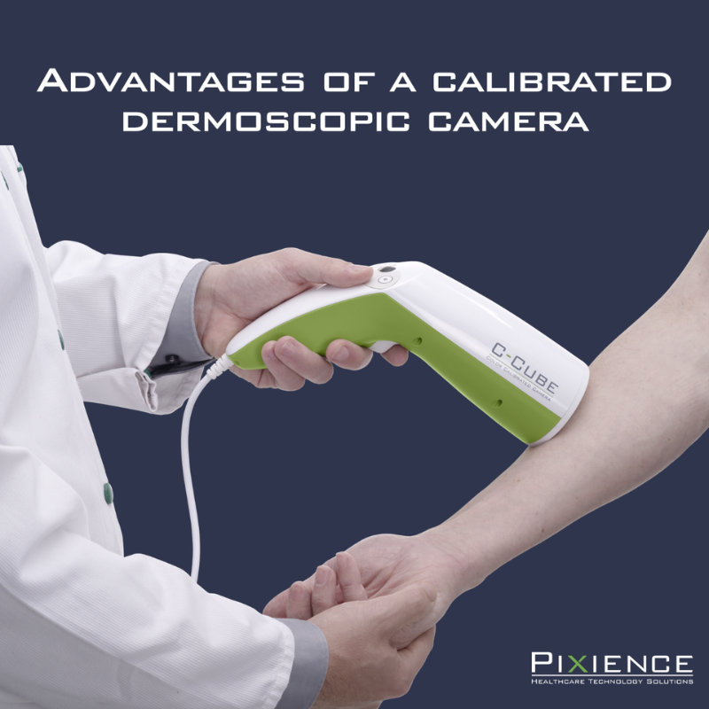
ARTICLE 3 – Why choose a calibrated dermoscopic camera?
In the field of dermocosmetic evaluation, imaging has become a central tool for objectifying the effectiveness of products. But with the diversity of solutions available, the question arises: how do you ensure reliable, repeatable, and actionable measurements?
Systems such as the C-Cube Clinical Research, based on calibrated microdermoscopic imaging, now provide a concrete response to these challenges by combining precision, standardization and quantitative analysis.
Standardization: a prerequisite for reliable data
One of the main challenges of clinical studies is the comparability of data over time, which is necessary to evaluate before/after evolutions.
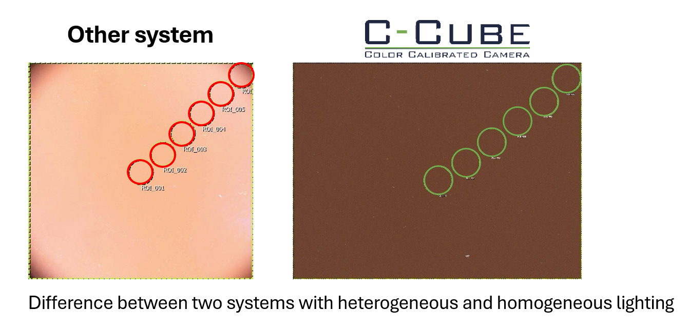
Without standardization, many biases can be introduced:
- Lighting variations,
- Differences in shooting angle,
- Influence of the operator.
These factors can alter the interpretation of the results, especially for subtle effects such as reducing redness or improving skin texture.
Calibrated dermoscopic cameras, such as the C-Cube, incorporate colorimetric and geometric calibration systems, ensuring:
- Faithful colour reproduction,
- Reproducibility of measurements over time,
- A homogeneity of the images over their entire surface.
This standardization ensures that the variations observed come from the product under test, and not from the measurement tool.
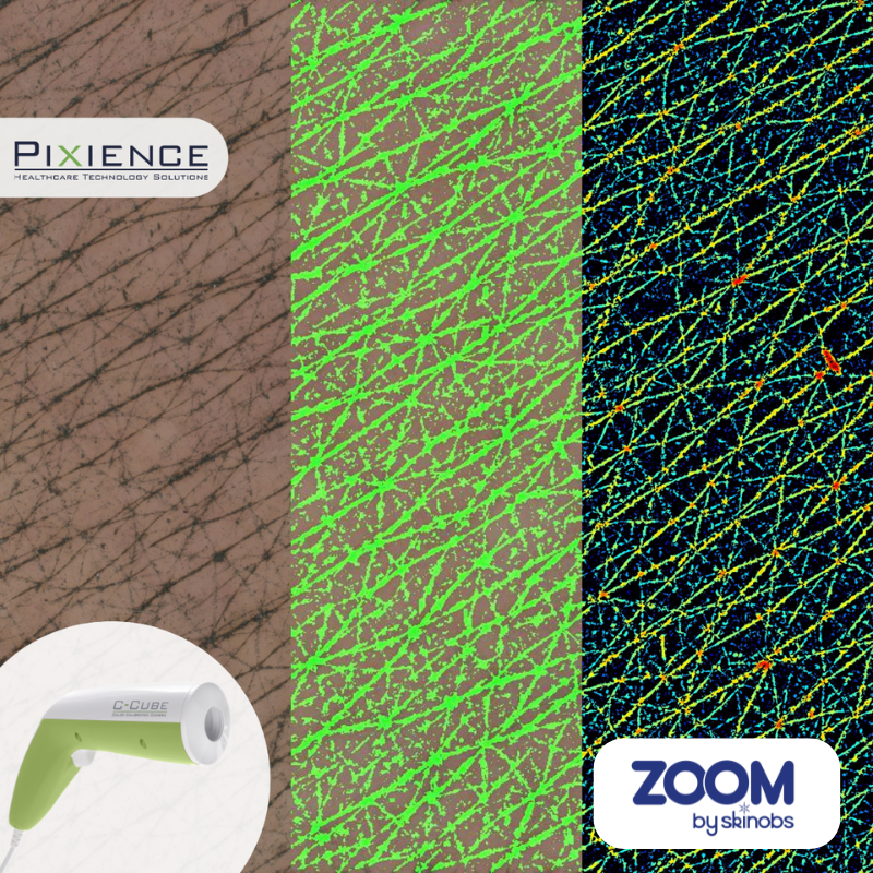
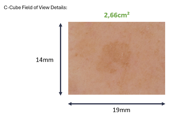
The microdermoscopic field: a scale adapted to efficacy studies
Beyond standardization, the level of detail of the image plays a decisive role.
Macro cameras allow a global vision, but quickly reach their limits when it comes to analyzing localized or subtle phenomena. Conversely, microdermoscopy offers a restricted but highly informative field of view, particularly suitable for clinical evaluation.
This type of imaging makes it possible to:
- Follow the evolution of a pigment spot,
- Analyze an inflammatory lesion,
- Observe the skin microrelief,
- Study specific hair areas.
By focusing on a targeted and reproducible area, the microdermoscopic field allows for precise longitudinal analysis, which is essential to demonstrate the efficacy of a product.
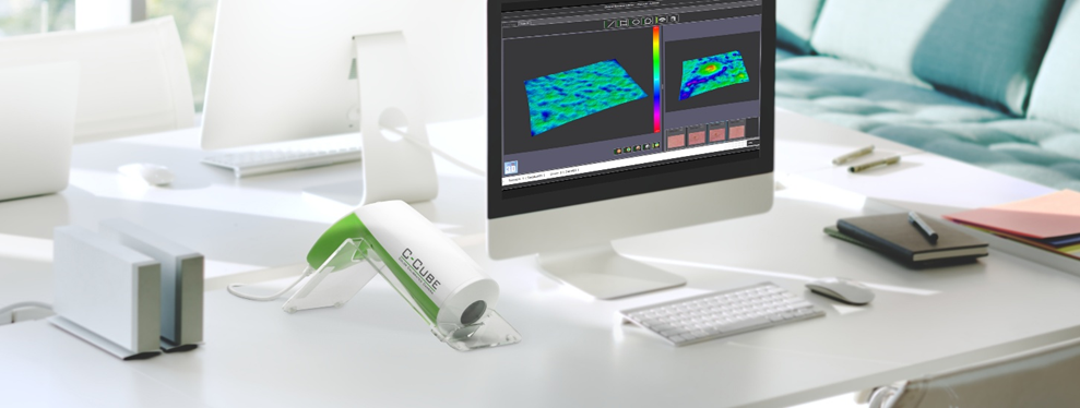
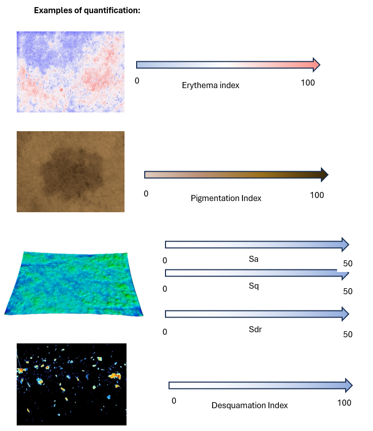
From image to data: the importance of quantification
Imagery alone is no longer enough. Today, the challenge is to transform the visual into measurable data.
Thanks to its analysis modules, the C-Cube allows to extract various quantitative parameters:
- Pigmentation and erythema indices,
- Roughness parameters (Sa, Sq, Sdr),
- Capillary measurements (density, diameter, length),
- Sebum or desquamation index.
This ability to combine visualization and quantification helps to strengthen the robustness of studies and effectively support claims.
Examples of quantification:
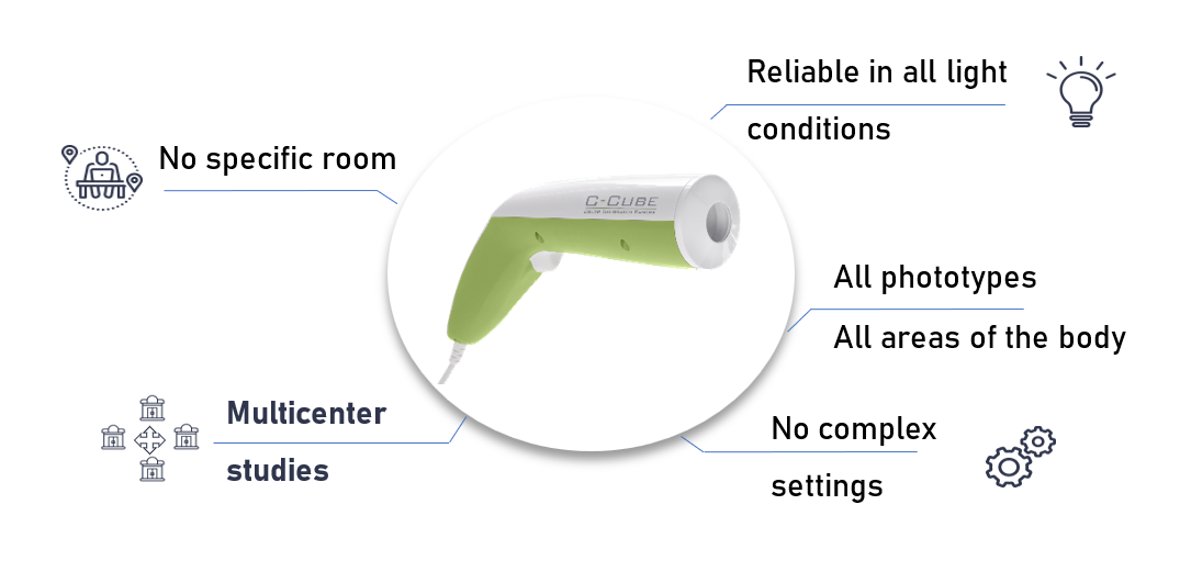
A technology adapted to clinical constraints
Beyond accuracy, the conditions of use are a key factor.
The C-Cube has been designed to be easily integrated into clinical protocols:
- Use without a dark room,
- Operator-independent reproducibility,
- Automated analytics,
- Compatibility with multicenter studies.
These elements facilitate the implementation of studies while maintaining a high level of scientific requirement.
A strategic choice for efficacy studies
Choosing a calibrated dermoscopic camera, such as the C-Cube, is not just a technological choice. It is a methodological lever for improving the quality of the data and the credibility of the results.
In a context where the requirements for validating claims are increasingly strong, having a tool capable of combining standardization, precision and quantification becomes essential.
Contact
Pixience
Sébastien Mangeruca
CEO
sales@pixience.com
https://www.pixience.com/c-cube-recherche-clinique/

To know more about Pixience expertise: https://www.skinobs.com/c/manufacturer.php?id=107